Labels of Lateral Flow Tests Home>>Labels of Lateral Flow Tests
Application
Contact Us
Labels of Lateral Flow Tests
List of materials used as a label in LFA is very vast which includes gold nanoparticles, colored latex beads, magnetic particles, carbon nanoparticles, selenium nanoparticles, silver nanoparticles, quantum dots, up converting phosphors, organic fluorophores, textile dyes, enzymes, liposomes and others. Any material that is used as a label should be detectable at very low concentrations and it should retain its properties upon conjugation with biorecognition molecules. This conjugation is also expected not to change features of biorecognition probes. Ease in conjugation with biomolecules and stability over longer period of time are desirable features for a good label. After the completion of assay, some labels generate direct signal (as color from gold colloidal) while others require additional steps to produce analytical signal (as enzymes produce detectable product upon reaction with suitable substrate). Hence the labels which give direct signal are preferable in LFA because of less time consumption and reduced procedure. Here we discuss some of the above mentioned labels in brief.
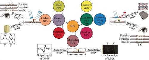
Figure 3-1 NPs Labels of Lateral Flow Tests
3.2.1. Gold nanoparticles
Colloidal gold nanoparticles are the most commonly used labels in LFA. Colloidal gold is inert and gives very perfect spherical particles. These particles have very high affinity toward biomolecules and can be easily functionalized. Optical properties of gold nanoparticles are dependent on size and shape. Size of particles can be tuned by use of suitable chemical additives. Their unique features include environment friendly preparation, high affinity toward proteins and biomolecules, enhanced stability, exceptionally higher values for charge transfer and good optical signaling. AuNPs can functionalize different biomolecules, including oligonucteotides and proteins. The antibodies bind tightly to its surface with noncovalent interactions, such as Van der Waals force and hydrophobic interactions. This way an immunocomplex is formed. The effects of using gold nanoparticle (AuNP) solution at various pH values and antibody concentrations are important to optimize the binding between antibodies and AuNPs.
The shape, size and stability of AuNPs are key parameters that affect the success of the LFA. Nano-colloidal gold with a diameter of 20 nm was used as detector reagent. AuNPs with the diameter larger than 60-70 nm are self-aggregated easily and unstable. The AuNPs/sample volume ratio directly affects the signal intensity of the test lines. A larger AuNPs/sample volume ratio provides more immuno-AuNPs conjugate to capture analytes in the sample, and thus a stronger signal is formed on the test lines of the strips. The particle size can be evaluated by spectral absorbance. There is a relation between size and maximum absorption wavelenght of visible spectrum of colloidal gold nanoparticles. After conjugation, the absorbance of peak increases and it shows that gold-antibody complex is formed. The stabilization of AuNPs-antibodies conjugate and the adsorption of various biomolecules on the membrane, sample, adsorbent and conjugate pads are important for eliminating matrix effect. In this context, bovine serum albumin (BSA) has been used as a stabilization agent of gold nanoparticles-antibody conjugate. Another advantage of BSA is that it avoids the matrix interference. Apart from BSA, other proteins, surfactants, polymers, and organic cations have been used as AuNPs stabilization agents in the labelled antibody solution and as a modifier of the nitrocellulose membrane. Normally, the results of an LFA are yes/no. For quantitative data from an LFA, it typically requires an image analyzer or comparision with a color scale card. To eliminate this need of using an additional device, barcode-style LFAs have been developed to provide rapidly a semi-quantitative result, based on relationship between the number of test lines appearing on the strip and the analyte concentration.
3.2.2. Fluorescent materials
Recently, in a number of studies, fluorescent nanoparticles are used rather than colorimetric markers and low detection limits are obtained.
3.2.2.1 Quantum dots
Quantum dots are ideal fluorescent labels and have been widely used to improve the detection sensitivity of LFA owing to their narrow emission spectra, broad excitation range and high fluorescent quantum yields. QDs have excellent brightness, size-tunable fluorescence emissions, large absorption coefficients, good stability, and high signal to noise ratio. The excellent of brightness and exceptional stability of QDs enable them to be detected and quantified sensitively. Water-soluble QDs with abundant carboxyl or amine groups coated in their surface provide them conjugate to proteins. The carboxyl groups of quantum dots are activated with 1-ethyl-3-(3-dimethylaminopropyl) carbodiimide (EDC) and sulfo-N-hydroxysuccinimide (NHS) chemistry, and the activated carboxyl groups react with amine groups on antibodies. These fascinating properties make QDs a robust label to develop sensitive LFA with capabilities to simultaneous by quantify multi analyte.
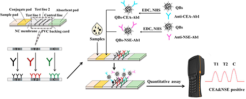
Figure 3-2 Illustration for Quantum-Dot Based Lateral Flow Strip Assays
For simultaneous detection of several compounds in a sample, two different formats of LFA have been developed. In the first format (as in Figure 3-2), LFA includes different lines for each analyte; in the second format, LFA includes one line for both analytes. In the first format, the different antibodies of analytes are immobilized in different lines. In the second format, all the antibodies are in the same line.
3.2.2.2 Lanthanide
The lanthanide chelate labels, such as Eu (III), Tb (III), Sm (III), and Dy (III), have been used in LFA to increase detection limit. The principle of this type LFAs is based on fluorogenic reaction. The size of lanthanide-labeled nanoparticles and AuNPs are quite close and therefore they are preferred as a label in LFA. The advantages of the lanthanide chelate based LFA is that it eliminates the background fluorescence. Thus, the sensitivity of LFA is increased. As compared to conventional fluorescent labels and quantum dots, lanthanide chelates possess distinctive and attractive fluorescent characteristics, such as long fluorescence lifetimes, narrow emission spectrum, large stokes shifts and outstanding photostabilities, all of which contribute to high assay sensitivity and accuracy. T lines and C lines were evaluated under UV light and the intensity of lines was read. The quantitative detection limit was determined by using strip reader. Eu (III) chelate microparticles were used as a label for ochratoxin A (OTA), prostate specific antigen (PSA), AFP, and cardiac troponin I (cTnI), and Pantoea stewartii subsp detection in LFA.
A fluorescent multiplex immunochromatographic strip was developed for the detection of three β-agonizts: clenbuterol, ractopamine, and sabuterol. The principle of this LFA was based on competition between analyte on the test line and analyte in the sample to bind fluorecence beads conjugated anti-analyte antibody. Each monoclonal antibody recognized its corresponding β-agonist in the conjugation pad. The unreacted antibodies were captured by β-agonist antigens immobilized on three lines in the nitrocellulose membrane reaction zone.
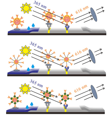
Figure 3-3 Illustration for Lanthanide (Eu III) Nanoparticles Based Lateral Flow Strip Assays
3.2.2.3 Up-converting phosphor (UCP)
Up-converting phosphor is a kind of lanthanide-containing, submicrometer-sized, ceramic particle, and its special composition and structure provide an excellent optical feature which can emit visible light when excited by infrared light. UCP particle is used as label to increase the sensitivity of LFA and is also used to detect hepatitisis B, Brucella, Schistosoma circulating anodic antigen, interferon-γ (IFN-ɣ), clenbuterol cTnI and PSA target-DNA.
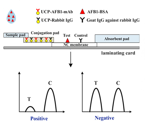
Figure 3-4 Illustration for UCP Based Lateral Flow Strip Assays
The main advantages of UCP reporters compared to other fluorescent labels are high sensitivity, long shelf life, permanent record (no fading), low matrix interference, and low cost. UCP decreases background interference and increases sensititvity, and this way a low LOD is achieved. UCP could be used to detect large molecules with sandwich assay and small molecules with competitive assay.
3.2.2.4 Other Fluorescent Materials
The use of fluorescent signals has attracted recent attention to enhance the detection signal and improve the sensitivity of LFA. Fluorescent microspheres (FMs), which have stable configuration and high fluorescence intensity, are multicolored and safe. Due to the lack of sensitivity when applying AuNPs, FMs are used mainly for the detection of several compounds, such as aflatoxin M1 (AFM1), tumor necrosis factor alpha (TNFα), procalcitonin, 4(5)-methylimidazole, E.coli. FMs bound to antibody faster and with more concentrations of antibodies. The increased antibody concentrations caused more interaction between E.coli and anti-E.coli antibody. Therefore low LOD was found.
Fluorochrome dye is used as a label in LFA systems. Fluorochrome dyes labeled antibodies have been used as dedector antibody for the construction of conjugation pad of LFA. Fluorescent nanosilica was used as a label for clenbuterol detection.
3.2.3 Carbon nanotube and Carbon nanoparticle
The carbon-based materials are used as a reporter in the development of LFA. They conjugate easily to biorecognition elements. Carbon nanotube, carbon nanoparticles, carbon nanostrings are the usual, preferred materials. Carbon nanotubes (CNTs) have gained significant attention since their discovery in 1991 due to their physical, chemical and electrical properties. Large surface area and good electrical and optical features make them suitable support material in biosensors and signal material in immunochromatographic strips. Researchers have developed a competitive lateral flow immunoassay for forchlorfenuron detection. The developed LFA is illustrated in Figure 3-5. In this LFA, black bands, a characteristic color of carbon nanoparticles, were observed. The carbon black-on-white signal allows for sensitivites down to the low picomolar range. Other advantages are very low cost, easiness of preparation, and the stability of the conjugates.
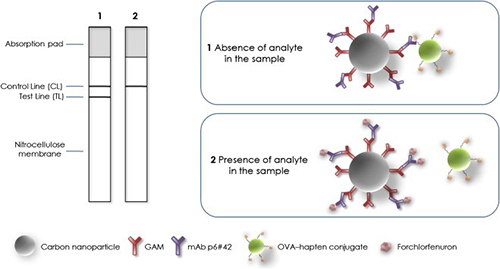
Figure 3-5 Illustration for Carbon Nanoparticle Based Lateral Flow Strip Assays
3.2.4 Superparamagnetic nanoparticles (MNPs)
Superparamagnetic nanoparticles (MNPs) are new labelling materials to develop a LFA. They increase the senstitivity almost 10 to 100 times. Additionaly, magnetic signals are produced by MNPs and they are stable over long periods of time. They have low background noise, because the magnetic material does not exist usually in the environment or in the tested samples.
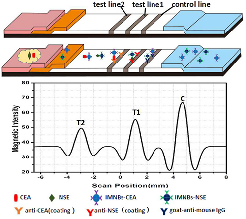
Figure 3-6 Illustration for MNPs Based Lateral Flow Strip Assays
Most of studies have showed that super-paramagnetic particles are new attractive material to construct a LFA, which will replace traditional labels due to its stable magnetic signal which can be entirely captured by devices, thus improving the sensitivity of LFA. The reasons for using these particles as label are its unique properties, such as magnetism in strong magnetic fields, and this property prevents aggregation and precipitation. A various LFA studies, which use supermagnetic particles as label, have been developed for Bacillus anthracis spores, paralbumin, Vibrio parahaemolyticus, cardiac troponin I, human chrorionic gonadotroponin (hCG) detection. The small size of magnetic nanoparticles (MNPs) enables short detection time. The increased magnetite content of MNPs provides stronger signal; also, the magnetite content has positive linear relationship with the signal intensity. The combination of superparamagnetic nanoparticles and antibodies are known as immunomagnetic beads (IMBs). Magnetic nanobeads contain Fe3O4 with antibody. IMB as a pretreatment material has been used to enrich and separate analyte by adding magnet. As a result, the analyte has been concentrated and matrix effect has been eliminated, and also the analytical sensitivity has been increased.
The signal from retention lines was visuliazed by naked eye and measured by portable magnetic assay readers. Colloidal gold particle is used widely as label and gold magnetic bifunctional nanobeads (GMBN) are a novel label. It possesses a Fe3O4/Au core/shell structure; thus, it can conjugate with biomolecules via non-covalent bonds. These particles separate the target bacteria from the samples by using magnetism and enrich these bacteria in the magnetic field. When GMBN based LFA was compared with traditional LFA, it was more sensitive and had shorter detection time.
3.2.5 Enzymes
In literature, three types of test strips have been used. The most common strip is based on capturing of analyte by an antibody labeled with colloidal gold, latex bead or fluorescent dyes to form antibody-antigen complex. This forming complex binds to other immobilized antibody on T lines and C lines. The second type of strip is based on chemical reactions which gives a color change. The third type of one is based on the enzyme-substrate reactions for color development. The formed color can be visualized by naked eye and standard color chart. This leads to imprecision caused by large variation in judgment due to human errors. Quantifications of signal by readers have been also developed, for example the refractometer for determining the optical density, the electrochemical signal for measuring the conductivity of metal labels and stripping voltammetric detection of metal ion labels. However, the requirements of expensive reading device make assay and costly less beneficial. To solve this problem, a non-instrumental approach has been developed where the color signal is presented in a ladder bar format.
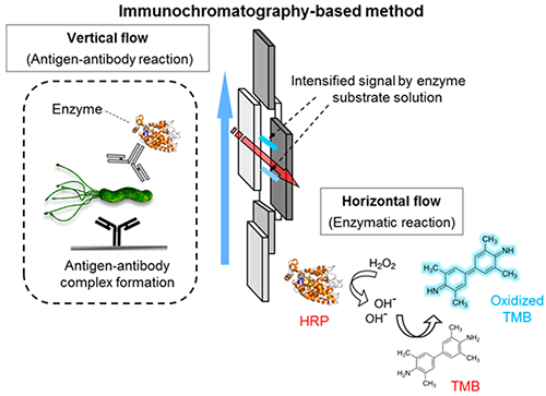
Figure 3-7 Illustration for Enzymes Based Lateral Flow Strip Assays
This strip was composed of four zones: a sample pad, peroxidase substrate zone, test zone, and absorbent pad. HRP was immobilized on test zone, and H2O2 and TMB (a chromogenic substrate) were consumed by immobilized HRP. A blue signal was seen after the reaction between HRP and H2O2. Another example of this type LFA was enzyme-catalyzed chemiluminescence based LFA. In this type of LFA, the antibody-antigen binding reactions and enzymatic reactions occurs simultenously. The enzymatic reaction generated C lines signal and the C lines emissions are measured by using a CCD camera.
To overcome the low analytical sensitivity as compared to the tradiotional enzyme assay, enzyme tracers have been used as an alternative to enhance the signal. AuNPs were combined with HRP and they found the detection limit as 0.01 pM target DNA without instrumention. An immuno-affinity test column assay based on enzyme based LFA was developed for chloramphenicaol detection. The gel coupled with CAP antibody was used as the test layer, and the gel coupled with CAP antibody was used as the control layer. This system principle was based on the competitive LFA format. With the help of HRP, the blue color appeared in negative samples and it disappeared in positive sample. The advantage of this system is that it is suitable for loading a large volume of liquid, which provides low detection limits.
3.2.6 Liposome
Liposomes are sphere-shaped artificial vesicles consisting of one or more phospholipid bilayers. Owing to their size and hydrophobic and hydrophilic character (besides biocompatibility), they have been particularly used in drug delivery. The properties of liposome differ considerably with lipid composition, surface charge, size, and the preparation method. They are very stable and their large internal volume provides interaction with several biorecognition elements, such as peptides, hormones, antibodies, sugars, and nucleic acids. These properties make them useful in field-portable or point-of-care sensor systems. The main disadvantages of liposomes are the structural weakness toward detergents, pH changes which an adverse effect on the ionic charges in the phospholipids bilayer, and osmotic pressure related swelling or crenation. Recently, lateral low assays in which liposome are used as label have been developed for Staphylococcal enterotoxin B (SEB), target DNA, allergenic peanut protein Ara h1 detection.
For the preparation of labels, a red dye marker, sulforhodamine B (SRB), is captured inside cavities of liposomes. Liposomes are able to encapsulate millions of dye molecules, which may thereby produce a very intense signal. This developed lateral flow assay principle is based on fluorescence measurement associated with the large amount of dye encapsulated into the liposome. The fluorescent signal is 15-fold better than with visual detection.
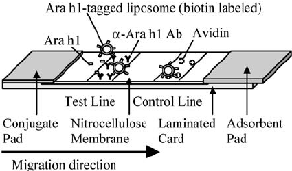
Figure 3-8 Illustration for Liposome Based Lateral Flow Strip Assays
In the lateral flow assays that were developed for allergenic peanut protein Ara h1, and maleimide-modified Ara h1 molecules were covalently conjugated to sulfhydryl groups on the liposome surface to form Ara h1-labeled liposomes used as the detector agent. Free Ara h1 molecules in the sample competed with Ara h1-labeled liposomes for binding to the limited number of anti-Ara h1 antibodies in the test line on the strips. The strip principle is based on competitive format and the obtained signal on the test line is inversely proportional to Ara h1.
USA
Tel:
Fax: 
Email: info@creativediagnostics.org
Add: 
Europe
Tel:
Email: info@creativediagnostics.org
Add: